The veterinary world is advancing at a rate of knots and the Animal Health Trust, Horse&Rider’s 2017 charity of the year, is where much of the magic happens. Lucy Turner visited to find out more
The veterinary world is advancing at a rate of knots and the Animal Health Trust, Horse&Rider’s 2017 charity of the year, is where much of the magic happens. Lucy Turner visited to find out more
When I think of equine charities, centres full of rescued, abused horses and ponies who need to find loving homes spring to mind. And while a huge part of equine charity focuses on horses and ponies in need, not all of it does. What I hadn’t considered was the amount of charity work that goes into making sure our horses stay happy, healthy and sound for years to come – that’s where the Animal Health Trust (AHT) comes in.
Specialist help
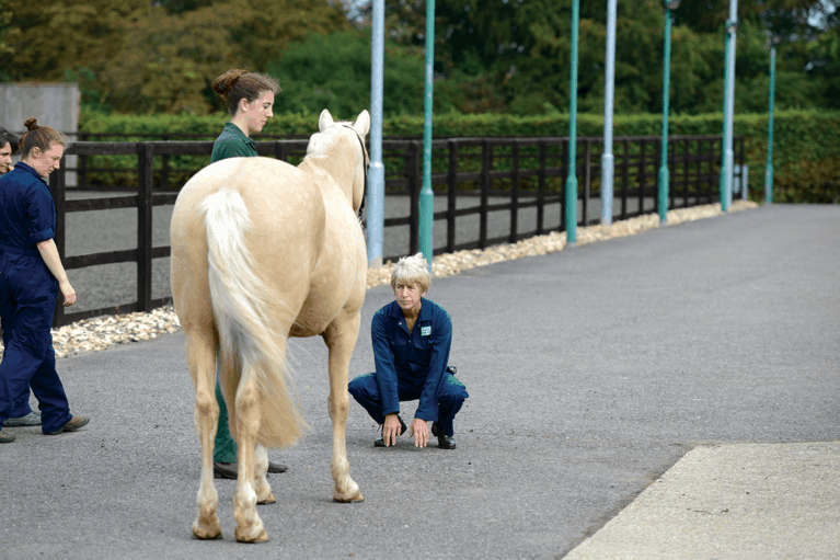
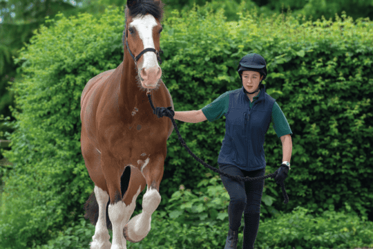
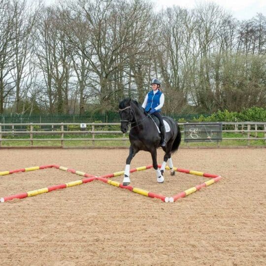
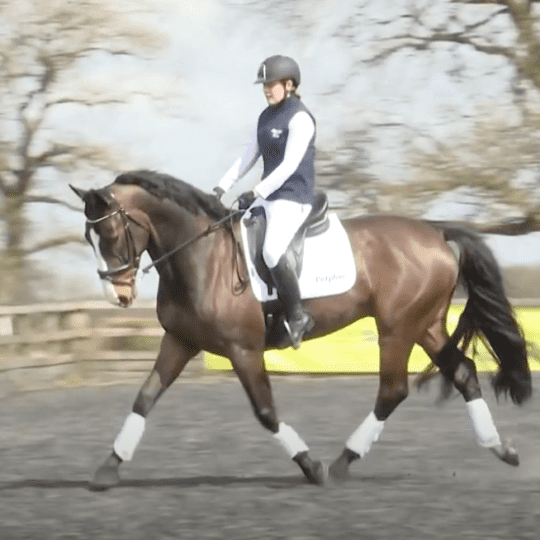
The Animal Health Trust has a referral clinic and the majority of the horses who come to the Trust are there for a lameness examination – with expert in orthopaedics, Sue Dyson, heading up this area of work, they couldn’t be in better hands. As horses arrived for their appointments and were led onto the yard by their owners, I couldn’t help but sense a hint of apprehension in the air. The girls on the yard welcomed them warmly and made the horses feel at home straight away, and the vets were kind and sensitive, as well as ultra-professional, but still there was a sense of unease.
It soon transpired that for many of the horses who come here, it’s a last chance saloon. Often they have ongoing lameness issues that, so far, no one has been able to resolve, so they are referred here by vets around the country in the hope that the cause and a solution can be found – I’ve often heard it said that if Sue Dyson can’t fix your horse, no one can!
The vets have state-of-the-art equipment at their disposal to help them reach a diagnosis, including a dummy rider for horses who aren’t safe to sit on, which allows the vets to see whether the horse is affected differently when he’s ridden. Once the area of pain has been located using nerve and joint blocks, the location is imaged using X-ray, ultrasound, bone scan and, in more complicated cases, MRI.
No imaging technique is 100% sensitive, so the vets use X-ray, ultrasound and bone scan to investigate problems, as they can all show up different things. For X-ray, there needs to be at least a 30% increase or decrease in bond density for the problem to be visible. And when vet, Laura Quiney, carried out some research on the sensitivity of bone scan, she discovered that in some horses, quite major bone changes weren’t picked up.
MRI detects almost all problems, but it’s incredibly expensive, so the vets try to get to the bottom of the problem using other techniques first. MRI is most commonly used for foot pain, because up to 80% of forelimb lamenesses are in the foot, and because it’s not possible to use ultrasound to image the tendons and ligaments in the foot due to the thickness of the hoof capsule.
Discover more about the ground-breaking work done by the Animal Health Trust in the March issue of Horse&Rider, in shops on 12 January, get your copy now
Your Comments
Related Articles
Newsletter Sign-up
Subscribe
Latest Issue

Brilliant Badminton
Added 22nd April, 2024
BRC secures SEIB sponsorship for further two years
Added 5th April, 2024
Tania Grantham’s diamond pole layout exercise
Added 27th October, 2022
Dressage to music – prelim level
Added 28th June, 2022