

Navicular disease has caused many horse owners heartache over the years, but vets have discovered that it may not be all it seems. Vet Lucy Meehan, from Langford Vets, explains
Our expert: Lucy Meehan BVSc MSc CertAVP(VDI) DipECVDI MRCVS is Senior Teaching Fellow in Equine Lameness at Langford Vets, University of Bristol. She is also an RCVS and European Veterinary Specialist in Diagnostic Imaging (Large Animals).
Over 90% of forelimb lamenesses in horses are located in the foot and, in the past, navicular disease was a common diagnosis, which filled horse owners with dread. The condition was career limiting at best and devastating at worst.
Because the navicular bone is small, buried deep inside the hoof and covered by a rigid hoof wall, it’s not the easiest structure for vets to investigate. However, now they have access to much better diagnostic equipment, it’s shed a whole new light on navicular disease.
The navicular bone
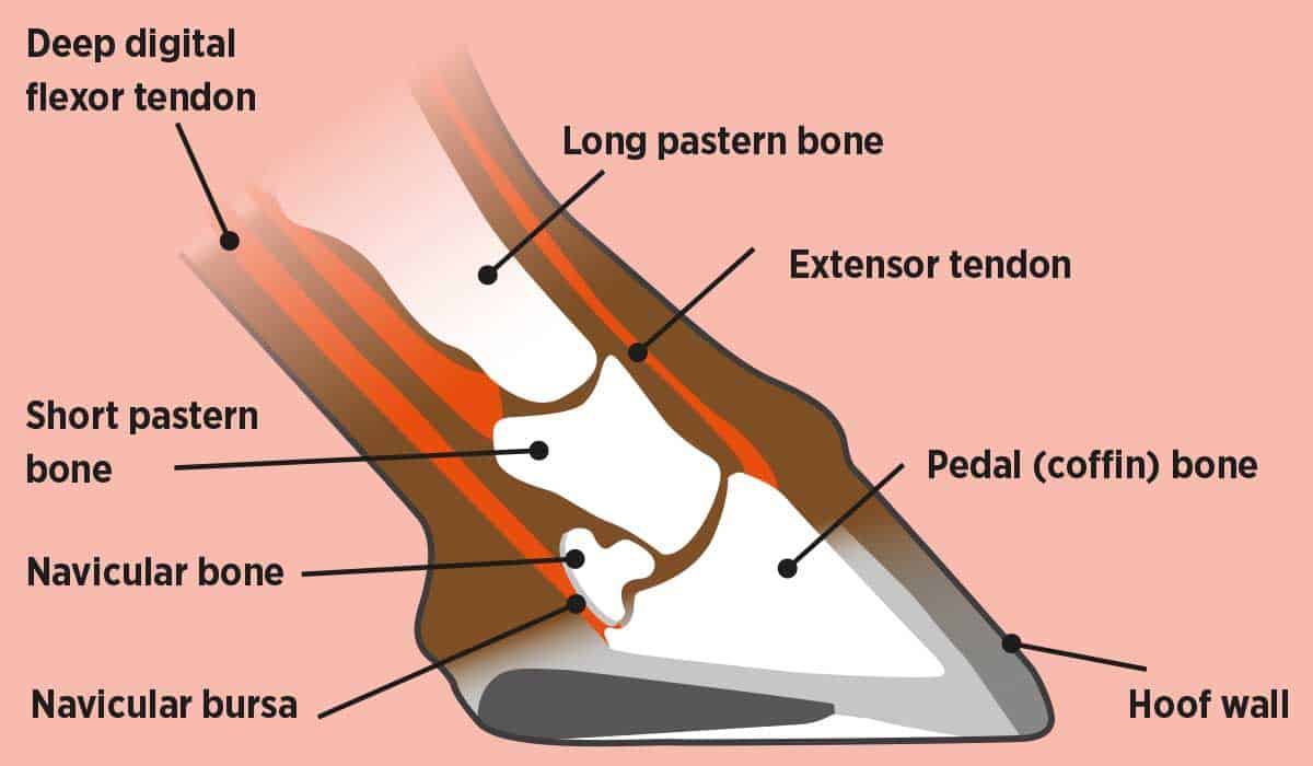
Let’s start with a quick anatomy refresher. The navicular bone is a small bone at the back of your horse’s foot and it’s held in place by a number of ligaments. It has a series of small channels, known as synovial invaginations, in one of its borders. These are holes in the bone that fill with synovial joint fluid and they’re entirely normal.
The navicular bone acts as a pivot for the deep digital flexor tendon where it attaches to the pedal bone, and the navicular bone and deep digital flexor tendon are separated by a small pocket of fluid known as the navicular bursa. The deep digital flexor tendon extends from above the knee, passing down the cannon bone to the foot. It’s responsible for flexing your horse’s foot and exerts pressure on the navicular bone with every stride.
Hoof conformation can change the forces exerted by the deep digital flexor tendon on the navicular bone and plays a part in the development of pain in this area. For example, navicular bone pressure will be greater in a horse who has long toes.
Is navicular a disease or a syndrome?
There’s also a condition known as navicular syndrome. It’s often thought to be the same thing as navicular disease, but there are important differences between the two…
navicular disease is a very specific diagnosis relating to pain that’s caused by disease of the navicular bone itself. The bone becomes more dense and there will be damage to the cartilage, which causes changes in the bone. Navicular disease is a degenerative disease, which means the changes will get worse with time
navicular syndrome, also known as palmar foot pain, is diagnosed when lameness has been located to the back half of the foot, but the cause of the pain hasn’t been confirmed. These horses have pain in the area of the navicular bone, however, in many cases they don’t have problems associated with the navicular bone, but with the surrounding structures
Navicular disease and syndrome are most common in the front limbs. The lameness can vary from shuffly, short strides in both front legs – which can lead to stumbling, reluctance to go forward, refusing fences and unwillingness to work – to severe lameness in just one front foot. The degree of lameness can vary day by day or can be consistent over a period of time.
How navicular is diagnosed in horses
It’s difficult to localise pain specifically to the navicular bone, as there are many structures closely related to it that can cause pain in this area. A diagnosis can usually be made following an examination and a combination of diagnostic techniques, including nerve blocks, X-rays and advanced imaging…
examination As with any lameness, an initial assessment is an essential part of the diagnosis. Your vet will look at your horse’s legs and spend some time feeling his limbs. They’ll also assess the conformation and appearance of his feet, and look at his shoeing if he’s shod. Then they’ll watch your horse walk and trot in a straight line, and on the lunge
nerve blocks If there’s a consistent lameness, nerve blocks will be carried out on your horse. This involves placing a small amount of local anaesthetic into nerves that go to a specific part of your horse’s leg or foot to numb them. Once the area has been numbed, your vet will watch your horse trot again. If his lameness improves, it means the pain has gone and the painful area has been pinpointed, although it will return once the anaesthetic has worn off
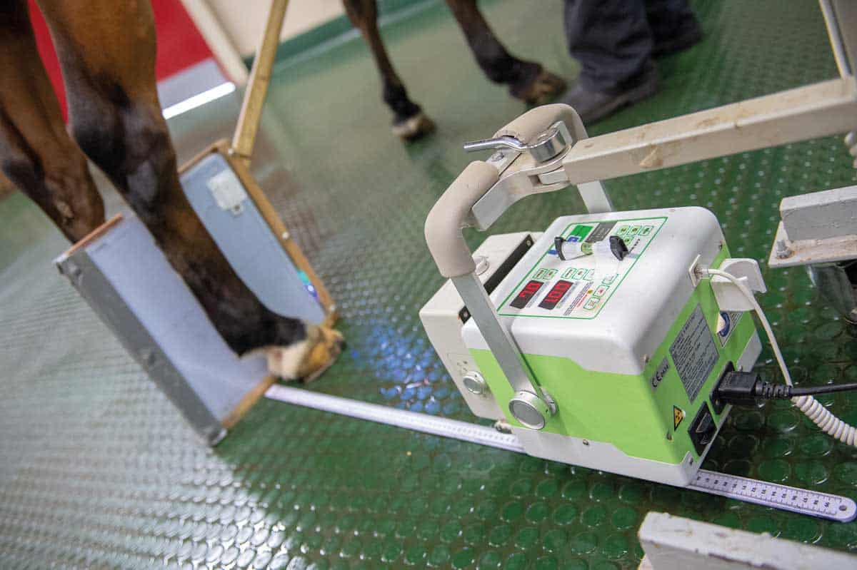
X-rays Usually, when lameness has been localised to the foot, X-rays are used to look at your horse’s navicular bone. Lots of factors will be assessed, but one of the most important signs of navicular disease is the number of synovial invaginations in the border of the navicular bone, and their size and shape – more, larger invaginations can be a sign of disease. Other signs include small bone fragments near the bone, changes in bone shape where the ligaments attach and changes in bone density. Horses can be diagnosed with navicular disease based on X-rays alone, but we now know that X-rays can underestimate the changes within the bone and they don’t enable us to look at the soft tissue structures surrounding it
Did you know?
Thanks to MRI, it’s become apparent that many horses who’d initially been diagnosed with navicular disease actually had injuries to surrounding structures that weren’t directly associated with the bone.
joint and bursa blocks Once nerve blocks have narrowed down the foot as the painful area, joint or bursa blocks can be used to try to localise the pain further. Your vet may inject local anaesthetic into the coffin joint, which numbs it and some of the surrounding structures. It’s also possible to inject into the navicular bursa, which is more specific for a diagnosis of navicular pain. However, it’s technically demanding and there’s a risk of introducing infection into the bursa or damaging the deep digital flexor tendon
advanced imaging Your vet can use computed tomography (CT) and magnetic resonance imaging (MRI) to view your horse’s foot as a 3D image, allowing them to see the bones and soft tissues in great detail. Recent advances have resulted in the development of an MRI machine that can be used with the horse under sedation rather than general anaesthetic, which has made it widely available and reasonably affordable. MRI will enable your vet to accurately diagnose the cause of your horse’s foot pain, whether it’s navicular syndrome, navicular disease, lesions in the deep digital flexor tendon, damage to the collateral ligaments of the coffin joint, coffin joint arthritis or bruising of the pedal bone
Achieving an accurate, specific diagnosis is important because there’s significant evidence to suggest that if your horse receives the correct treatment for his injury soon after the onset of lameness, he has a much higher chance of recovering and returning to work. As there are a number of different conditions that cause pain in the same area of the foot, all requiring slightly different treatment, it’s essential to use MRI to understand exactly what’s going on – all these injuries are impossible to diagnose with X-rays alone.
Treating navicular
Specific treatment options will depend on the cause of the pain in the navicular area, but there are common themes to treatment for most of the conditions…
remedial farriery Poor foot balance is surprisingly common in horses with foot pain. It’s impossible to accurately determine where the pedal bone is within the hoof capsule without X-rays, so it’s likely your vet will want to work with your farrier to improve your horse’s foot balance. For horses with confirmed navicular disease, additional heel support, heel elevation or placement of a rolled toe shoe may help
rest Many conditions require a period of rest. As a general rule, soft tissue injuries, such as deep digital flexor tendon tears and collateral ligament injuries, require prolonged periods of box rest with controlled exercise, whereas bony injuries, such as navicular disease or coffin joint arthritis, may require a more active recovery programme
medication Depending on which structures are involved, it may be necessary to treat the coffin joint or navicular bursa. This usually involves injecting a highly concentrated steroid anti-inflammatory directly into the structure. In addition, a drug called hyaluronic acid may be used. This is a normal component of the fluid within the joint or bursa and it reduces in quality when there’s an injury. Injecting into the joint or bursa helps to restore it to a more normal state
Prognosis
The prognosis will depend on the cause of the pain. Horses with navicular disease, coffin joint arthritis or other bony changes often improve with treatment, but many of these changes are degenerative in nature, meaning they’ll get worse with time, even with good treatment and management. For deep digital flexor tendon injuries, the prognosis varies depending on the location of the injury, the extent of the damage and the response to treatment. However, they can be very difficult to manage. Injuries to the collateral ligaments of the coffin joint often respond well to treatment, but may require a prolonged period of rest (8–12 months) and controlled exercise.
Advanced imaging and its widespread availability has meant that horses who would previously have been diagnosed with navicular disease may now be diagnosed with other injuries within the hoof capsule. This means they can be more accurately treated and they stand a much better chance of returning to work. But interestingly, it turns out that navicular disease is much less common than previously thought.
With thanks to Langford Vets for their help with this feature, langfordvets.co.uk
Your Comments
Related Articles
Newsletter Sign-up
Subscribe

Brilliant Badminton
Added 22nd April, 2024
BRC secures SEIB sponsorship for further two years
Added 5th April, 2024