
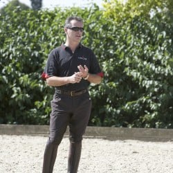

If your horse goes lame, diagnostic technologies will allow him the most appropriate treatment possible, as vet Jonathan Anderson explains
Lameness is one of the most common reasons for calling your vet and a condition that affects most horses at some point. Sometimes the cause of the lameness is known, or immediately obvious. At other times it isn’t as clear-cut, especially if more than one limb is involved.
There can be a multitude of reasons why your horse is unsound. If you think your horse is lame and it’s not an emergency, the first thing to do is to check for anything you might be able to deal with yourself, such as a stone in the hoof or a loose shoe. Is there heat in any of his limbs or are there signs of a recent injury, such as a cut or swelling, no matter how small or seemingly insignificant? These are important checks, as they may be crucial to knowing why your horse is lame.
Initial checks
The next step is to call your vet to discuss your findings, or what may have happened to make your horse lame. If you book a call-out, your vet will repeat your initial checks. They’ll examine your horse’s feet, see if there’s any response from hoof testers and check your horse’s digital pulses. If raised, this can point towards foot-related issues such as laminitis, an abscess or bruising.
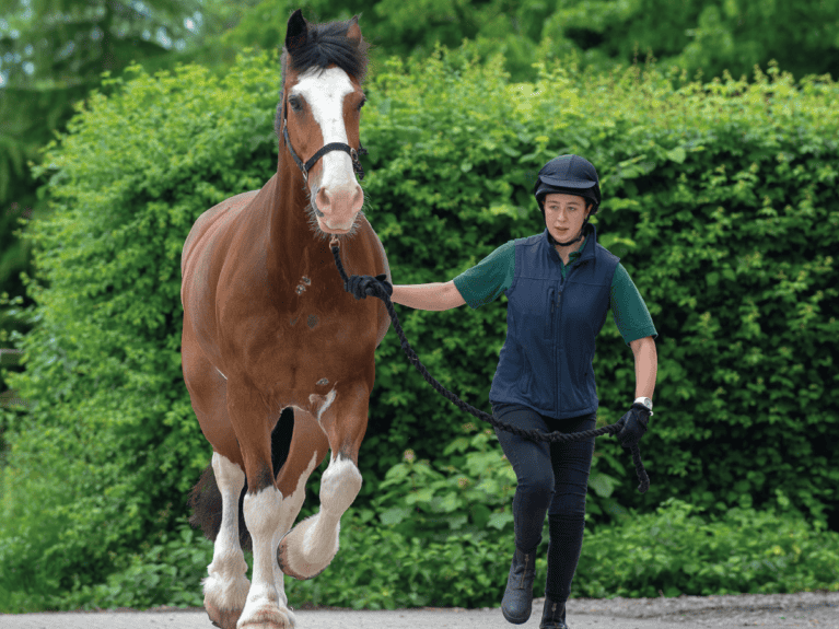
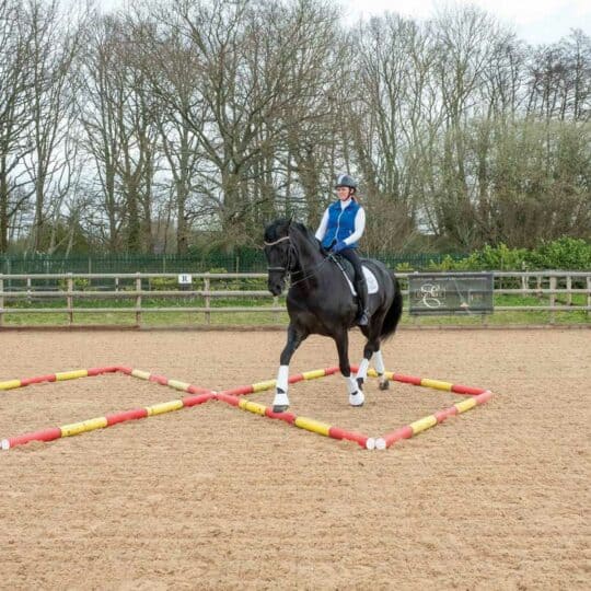
Your vet will ask you to walk and trot up your horse in a straight line away from and towards them. They may perform flexion tests – when the leg is held in a flexed position for 30–60 seconds before the horse is trotted away. These checks aren’t an exact science, but they can help identify the part of the leg to focus on.
Your vet may also watch your horse on the lunge, as lameness can be more evident on a circle. It’s useful to do this on a firm, non-slippery surface (in walk and trot) and also a soft surface (in trot and canter), on both reins.
They may also palpate your horse’s neck, back and sacroiliac region, because lameness can be caused by pain in other areas of the body.
Saddle up
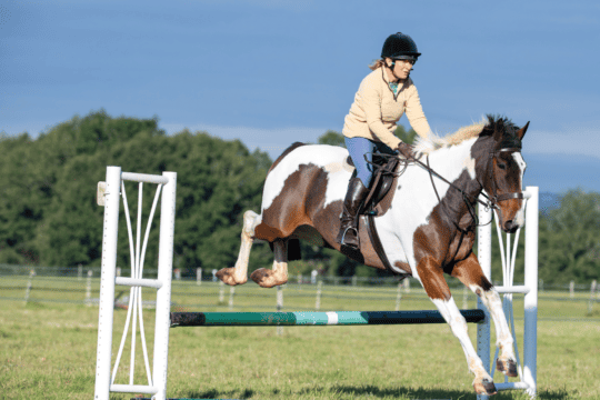
Your vet may ask you to ride your horse to see if there are any changes when he’s under saddle. In cases where you’re able to feel the issue more than see it, the ridden evaluation allows your vet to observe what you’re feeling while you give useful feedback from the saddle.
A closer look
If after the initial assessment it’s still not clear why your horse is lame, your vet will discuss any options with you, which is likely to be further diagnostic tests.
If the lame leg is known, but the specific part affected is not, nerve blocks are often the next step. This involves numbing specific sections of the leg by injecting local anaesthetic around a nerve or nerves, or into a joint or tendon sheath. The horse is then trotted up again – if he’s then sound, the area that has been blocked is the culprit.
Many nerve blocks can be done at your yard, but if multiple blocks are needed, it’s more time-effective for them to be done in a hospital or practice environment, especially those that require ultrasound or other imaging techniques to ensure accurate placement.
A case for referral
If, after your horse’s initial lameness examination, more advanced diagnostic testing is required, your vet may refer him to a hospital with the appropriate facilities. There, your horse will be examined by specialist vets who are highly experienced in investigating lameness and performance issues.
Referral hospitals provide the best conditions for your horse to be thoroughly examined. Team members will trot up and lunge your horse, and possibly ride him, and you will be able to stand with the vet while they draw their conclusions.
Top tip
Always trot up your horse on a loose rein to enable freedom of movement – particularly in his head.
Gait analysis
Technology is invaluable when assessing lameness, because it removes human subjectivity. Objective gait analysis can identify deficits in movement so slight that even the well-trained eye can’t see them. This is particularly useful if the lameness is very subtle, the horse appears unsound in more than one leg, or for establishing changes in lameness patterns following blocking.
Sensors are placed at the poll, withers and hips to measure stride length and differences in symmetry as each leg lands. Lifting the head whenever the lame limb touches the ground is the classic sign of foreleg lameness. Poll sensors can detect a 5mm difference in symmetry, compared with 20mm seen by the human eye. In hind limbs, the sensors measure a drop of the hip or a tilt in the pelvis.
Turning to technology
Modern equine hospitals are equipped with advanced diagnostic equipment and these tools are often used together so scans and images can be compared to make the most accurate diagnosis…
- digital X-ray is commonly used in lameness investigations to look at the joints and bones of the leg as well as the skull, neck and back
- ultrasound is used if the lameness involves tendons, ligaments or soft tissues not visible with digital X-ray. High-frequency sound waves pass through the body before bouncing back to create live images that can be viewed on a computer screen
- magnetic resonance imaging (MRI) creates highly detailed images of bone and soft tissue. MRI is often used to assess horses with issues in their feet, but it can also be used higher up the leg
- computed tomography (CT) is a rotating X-ray machine that produces superbly detailed 3D images of bones and joints. Your horse can stand for the fetlock and below, but for other areas, he’ll be anaesthetised to prevent any movement
- nuclear scintigraphy, or bone scanning, spots areas of inflammation, which is useful if multiple problems are contributing to lameness. A radioactive marker is injected and any inflammation shows as hot spots that are then captured by a gamma camera
Diagnosis and treatment
Thanks to the sophisticated equipment now available, vets can accurately diagnose most causes of lameness or poor performance. Armed with this information, they can advise on the prognosis for your horse and recommend appropriate treatment and management strategies, which could involve medication, surgery, rehabilitation or remedial farriery.
Did you know?
When radioactive substances are used, strict safety procedures must be followed and your horse will stay at the equine hospital for up to 48 hours, until the radioactivity has cleared from his system.
The road to recovery
Once all the tests are concluded, any findings will be explained to you and your horse’s usual vet, who will also receive copies of the diagnostic images.
A suitable treatment programme can then be put in place, and this is likely to be a team effort involving yourself, your vet, the referring vet, your farrier and your vet physio.
In this way, with everyone working together, you can set your horse off on the right road to recovery – one that leads back to soundness.
Did you know?
MRI scans can help detect early signs of bone disease and fractures that aren’t visible on X-rays.
Our expert: Jonathan Anderson is Clinical Director and Surgeon at Rainbow Equine Hospital in Yorkshire, and an FEI veterinary delegate for eventing.
Your Comments
Related Articles

Newsletter Sign-up

Subscribe

Latest Issue

Brilliant Badminton
Added 22nd April, 2024

BRC secures SEIB sponsorship for further two years
Added 5th April, 2024


Tania Grantham’s diamond pole layout exercise
Added 27th October, 2022

Dressage to music – prelim level
Added 28th June, 2022